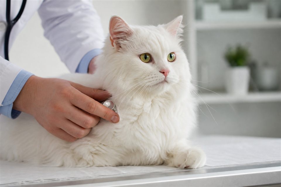
Oliver's cat, Fig, had always looked slightly sleepy — but when Priya noticed a pale pink membrane covering the inner corner of his left eye while the right eye looked normal, she knew this was different. The third eyelid, or nictitating membrane, had slid partway across the globe and stayed there. Fig was eating and playing, yet that one eye looked half-closed behind a fleshy curtain. Priya read about "haw syndrome" and wondered if it was harmless or a sign of something deeper going on elsewhere in his body.
The third eyelid is a normal feline structure — a T-shaped cartilage frame covered by conjunctiva that sweeps across the eye for protection and tear distribution. Healthy cats keep it hidden except during sleep or brief blinking. Persistent protrusion in one eye, both eyes, or alternating eyes signals an underlying trigger: ocular pain, nerve dysfunction, dehydration, gastrointestinal illness, or idiopathic haw syndrome where the membrane elevates without obvious cause after a viral or inflammatory episode.
Key points at a glance
- The third eyelid showing in one eye can reflect local eye disease, nerve issues, or systemic illness — not only an eye problem.
- Haw syndrome ( bilateral third eyelid protrusion) often follows gastrointestinal upset or viral infection and may resolve on its own over weeks.
- Unilateral protrusion warrants a thorough eye exam — corneal ulcer, glaucoma, and retrobulbar abscess can all trigger it.
- Other causes include dehydration, weight loss, tetanus-like toxins, and Horner's syndrome affecting nerve supply to the eye.
- Persistent third eyelid elevation beyond two to three weeks, or any eye pain signs, needs veterinary evaluation.
What the third eyelid does and why it becomes visible
Cats have upper and lower eyelids like humans plus this third protective layer at the medial canthus — the inner corner near the nose. Smooth muscle and neurologic input control its position. When the eye hurts, when sympathetic nerve tone drops, or when systemic illness reduces normal muscle tension, the membrane elevates and stays visible — the "haw" of haw syndrome.
Owners describe a white or pink film crossing the eye, sometimes both eyes, sometimes one. Fig's left-only presentation points Priya's vet toward localized causes first: something affecting that eye specifically rather than a whole-body GI trigger that often hits both sides symmetrically.
Haw syndrome explained
Haw syndrome — also called bilateral third eyelid protrusion with no other ocular signs — is a diagnosis of exclusion. It typically appears in young cats after:
Gastrointestinal inflammation — Diarrhea, vomiting, or inflammatory bowel flare-ups.
Respiratory viral infection — Calicivirus and herpesvirus associations appear in case reports.
Parasitic or protozoal illness — Less common but documented.
No identifiable trigger — Some cases remain idiopathic.
The membrane elevation is thought to involve disrupted neurologic signaling between the gut and eye — possibly via the autonomic nervous system — rather than direct eye damage. Cats often feel mildly unwell or recently recovered from GI signs when haw appears. Crucially, the eyes themselves usually lack ulceration, discharge, or pressure elevation on exam.
Prognosis is generally good: many cases resolve spontaneously over days to several weeks as the underlying illness clears. Supportive care and treating GI disease if present speed recovery. Fig only has one eye affected, so haw syndrome in the classic bilateral sense is less likely — Priya's vet will still ask about recent diarrhea or viral symptoms.
Unilateral third eyelid protrusion — local causes to rule out
When one eye alone shows the membrane, investigate that eye first:
Corneal ulcer or abrasion — Pain causes third eyelid elevation; squinting, tearing, and light sensitivity accompany it.
Glaucoma — Elevated intraocular pressure; eye may look cloudy or enlarged; painful and vision-threatening.
Uveitis — Inflammation inside the eye; may follow infection or immune disease.
Conjunctivitis — Discharge and redness with membrane involvement.
Foreign body — Plant material under the third eyelid.
Retrobulbar abscess or tumor — Pressure behind the eye pushes the globe forward and elevates the membrane.
Horner's syndrome — Nerve damage causing third eyelid rise, small pupil, and sunken eye on the same side; causes include ear infections, trauma, and neck injuries.
Priya should look for discharge, cloudiness, different pupil sizes, or Fig rubbing the left eye. Any of those push urgency.
Systemic and neurologic causes affecting one or both eyes
Dehydration and severe weight loss — Loss of orbital fat lets the eye sink slightly, making the third eyelid more visible in both eyes; common in chronically ill senior cats.
Tetanus — Rare in cats but causes rigid third eyelid elevation among other signs.
Dysautonomia — Severe autonomic nerve dysfunction; third eyelid protrusion with dry nose, dilated pupils, and GI stasis.
Polycystic kidney disease or other chronic illness — Indirect effects through debilitation.
Blood work, eye pressure measurement (tonometry), fluorescein stain for ulcers, and detailed neurologic exam help sort these categories. Fig's normal appetite and energy are reassuring but do not replace an eye exam — glaucoma and ulcers can hide behind subtle signs early.
What Priya should do next
Schedule a veterinary appointment within a few days for unilateral third eyelid protrusion — sooner if Fig squints, the eye looks cloudy, or discharge appears. Note recent vomiting, diarrhea, upper respiratory symptoms, weight change, and any trauma to the head or neck.
At home, prevent Fig from pawing the eye; use an Elizabethan collar if self-trauma starts. Do not use human eye drops left over from another pet unless prescribed — wrong steroids on an ulcer worsen rupture risk.
If both eyes suddenly elevate after a GI episode and Fig otherwise acts normal, many vets adopt a watchful approach with recheck in two weeks — but only after confirming the eyes themselves are healthy on exam. Unilateral cases rarely fit pure haw and need local investigation.
The bottom line
A third eyelid showing in one eye means Fig's body is signaling something — eye pain, nerve disruption, or occasionally a post-viral haw pattern more typical when both eyes are involved. Priya noticed a real change from Fig's usual sleepy look, and unilateral protrusion deserves a slit-lamp exam and pressure check before assuming it will pass. Haw syndrome in the classic sense is a bilateral, often post-GI finding that usually heals with time; one eye alone points the vet toward corneal injury, glaucoma, or Horner's syndrome first. The membrane is the symptom — finding what raised it is the job.
This article is for general information only and is not a substitute for advice from your veterinarian.